#microscopy

waynerad@diasp.org

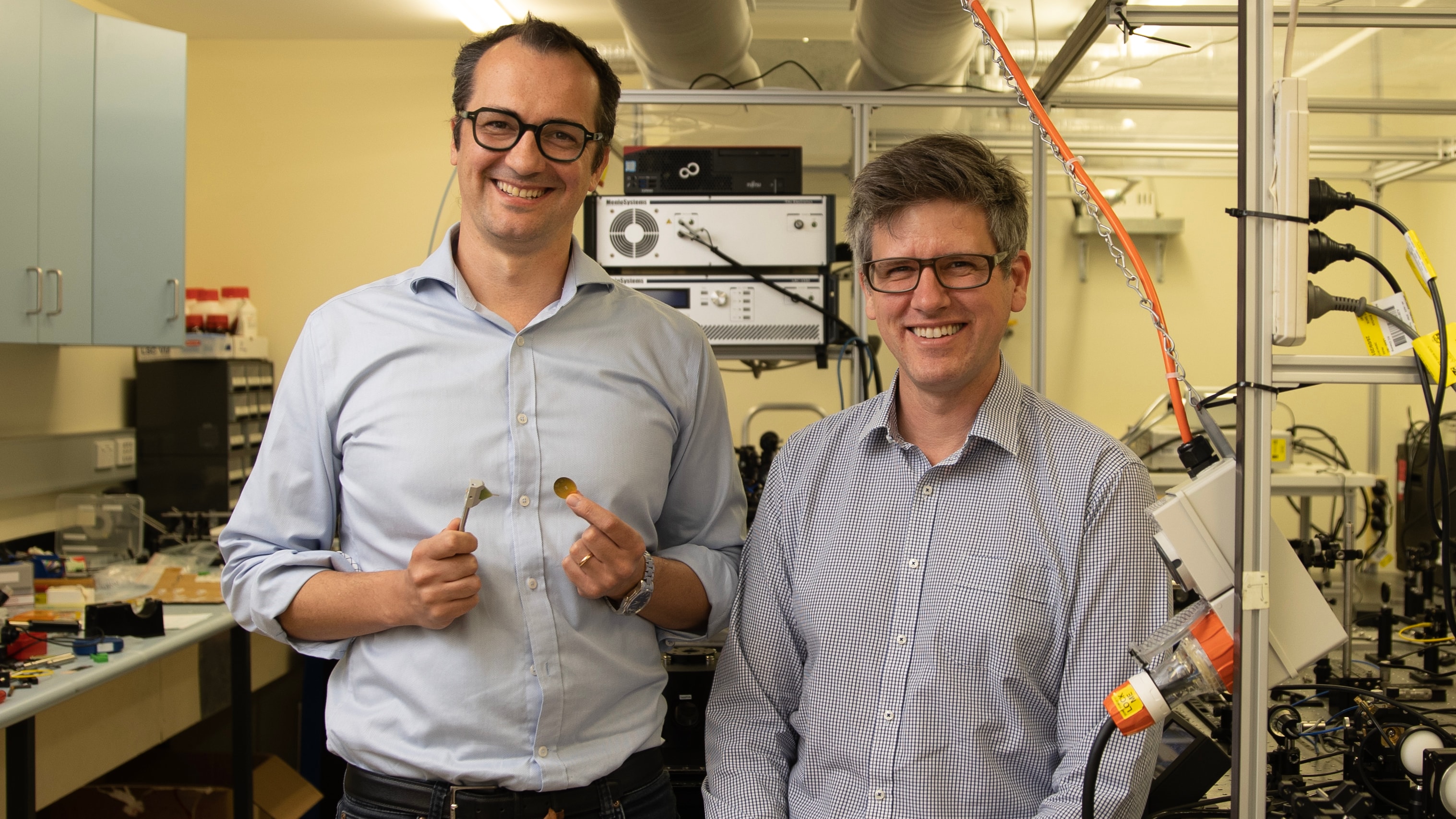
OpenFlexure "uses 3D printers and off the shelf components to build open-source, lab-grade microscopes for a fraction of traditional prices. Used in over 50 countries and every continent, the project aims to enable Microscopy for Everyone."

"An open flexure microscope is built from a combination of off-the-shelf electronics, standard optical equipment, and 3D printed parts. The 3D printed parts are designed to be made on any entry grade printer anywhere in the world." "Nothing is proprietary or hidden."

"The finished microscope can run automatically for several hours, scanning samples with a built-in autofocus. The 8-megapixel camera is comparable to many commercial sight scanners, achieving a resolution below 400 nanometers."

"In practical terms this means that individual cell damage or parasites can be identified on a microscope with parts costing under $300. The stage is fully automated, intelligently planning its own path around samples. It can also self-calibrate, warning the user if there's any damage that could impact the diagnosis. The automated stage allows huge data sets to be collected and stored.

"In pathology, this let samples be archived, shared, or used for the training of medical students. this can also be the platform for low resource artificial intelligence systems or automated image processing, making emerging technologies more accessible in low resource settings."

Something else I didn't know exists until just now. Developed at the University of Bath, University of Cambridge, and the University of Glasgow, with contributions from the Baylor College of Medicine, Bongo Tech & Research Labs, and Mboalab.

The OpenFlexure Project

#solidstatelife #3dprinting #microscopy

ramnath@nerdpol.ch

#RebeccaMarina ¦ #Cymatics and #sound #therapy, " #darkfield" #microscopy, and #prayer

https://x.com/BrianRoemmele/status/1854969616482943247

In this fascinating video clip shared by Brian Roemelle on X we witness what he describes as the following:
"Dr. Rebecca Marina had her red blood cells studied under different emotions and states.

When she felt sad, her blood cells actually became tear-shaped!
But when she felt #love, her cells turned back to normal and even #sparkled with tiny #lights.

Oh get ready for the #woo woo. When scientists prayed over her blood cells (separated from her body for hours), they started glowing and pulsing at the same rate as her heart.

The cells that weren't prayed over?

No movement at all."

We've a really good repository on the forum of the effects of sound on healing of course but seeing a clip of the effects of prayer biologically is really very remarkable indeed.

"This is because emotions are amphipathic peptides, which bind to the lipid bilayer of red blood cells leading to agglutination (clumping).

Praying at a distance helps due to loving intention communicated via biophotons, transmitting oxytocin release, which relaxes blood vessels increasing red blood cell mobility.

All cells in our body release and communicate with others cells via biophotons at the speed of light, just like the internet."

https://digitalrepository.unm.edu/cgi/viewcontent.cgi?article=1787&context=math_fsp&t&utm_source=perplexity

waynerad@diasp.org

"Why Recursion Pharmaceuticals abandoned cell painting for brightfield imaging."

"The bet of the whole company is the following: take a drug (either from a chemical screening library or dreamt up by a model), wash it over a plate of cells, visually observe how the cells react to it, and repeat this a few billion times across many different cells + many genetic knockouts of those cells (which acts as a model of a disease) + many different drugs. The whole idea is also known as phenomics -- the study of the visual phenotype of organisms. In this case, the 'organism' is a small set of cells. Once you've done that, train a model on those petabytes of cellular images, along with the genetic or chemical perturbation applied."

"The Recursion bet is that the resulting model would acquire an extremely strong understanding of the interaction between the visual morphology of cells, their genetic makeup, and drugs applied to them -- predicting the rest given one (or two) of the others."

"But this essay isn't about Recursion's drug discovery strategy." "This essay is about how Recursion takes pictures of cells in the first place, why it (officially) changed its approach just a few months ago, and why I think the decision to change it is a lot more interesting than people think."

"The superiority of using raw, unaltered cell painting images as input over hand-crafted features was established by 2019, but, as time went on, the utility of cell painting at all also became suspect. Per a 2022 Scientific Reports paper, one could nearly perfectly predict what the cell painting image would be given a brightfield image."

The article explains that "brightfield" microscopy was what was originally just "looking through a microscope": you put the cell you want to observe on a slide and shine bright light on it from behind and look at it through the microscope. This in contrast with "cell painting" where dyes are used to differentiate between RNA, endoplasmic reticulum, mitochondria, cytoskeleton/cell-membrane, and the cell nucleus.

"Another example of the bitter lesson; pre-imposing structure on your data is useful for a human, but detrimental to a machine."

"But there is another side benefit of going the brightfield route: the ability to do time-lapse microscopy."

Why Recursion Pharmaceuticals abandoned cell painting for brightfield imaging

#solidstatelife #biotech #microscopy

waynerad@diasp.org

Transparent mice.

"The addition of common dye molecules that absorb in the near ultraviolet and blue regions improve optical transparency in nearby longer wavelengths. In essence, by causing sharp absorption in the blue region, the refractive index in the red part of the spectrum is increased without increasing absorption."

"By applying the Lorentz oscillator model for the dielectric properties of tissue components and absorbing molecules, we predicted that dye molecules with sharp absorption resonances in the near-ultraviolet spectrum (300 to 400 nm) and blue region of the visible spectrum (400 to 500 nm) are effective in raising the real part of the refractive index of the aqueous medium at longer wavelengths when dissolved in water, which is in agreement with the Kramers-Kronig relations. As a result, water-soluble dyes can effectively reduce the refractive index contrast between water and lipids, leading to optical transparency of live biological tissues."

The dye molecule they used? Tartrazine. Also known as "Yellow 5". Used in Doritos.

"So far, the scientists behind the new discovery have used the method to see the organs in a mouse's intact abdomen, glimpse the pulsing vessels surrounding a rodent skull, and to get an exceptionally clear view of muscle tissue through a microscope."

The dye in Doritos can make mice transparent | Popular Science

#discoveries #biology #biotech #microscopy

waynerad@diasp.org

"Superlensing without a superlens: microscopes boosted beyond limits".

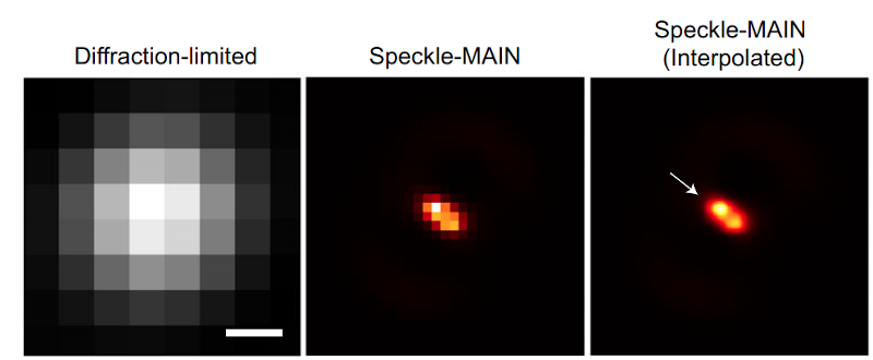
So what this is about is, in light microscopes, the limit of resolution is half the wavelength of light used. Fine details cannot be seen below this limit. This technique breaks through the diffraction limit by a factor of nearly four times. This has the potential to greatly enhance biomedical imaging.

There is special interest in terahertz frequencies for biomedical imaging because light at terahertz frequencies has the ability to penetrate tissues deeper than visible light. It interacts differently with water and various biological materials, enabling images with good contrast. It is non-ionizing, which means it doesn't harm the tissues it penetrates. If hooked up to a spectrometer, it can be broken out into a spectrum and specific molecules identified. But it has a longer wavelength than visible light, so the diffraction limit is a problem.

This brings us to the virtual superlensing effect. The key to understanding this is to think of there being two types of light waves reflected off the object: propagating waves and evanescent waves. The "propagating waves" are what you normally think of as light waves and are what you use to see the object -- but you can only see features that are above the diffraction limit. The "evanescent waves" are the waves that carry the fine detail information, but they decay exponentially as you move away from the object under observation. The virtual superlens, however, is a clever way to capture the information carried by these "evanescent waves". The system uses Fourier expansions to mathematically "reverse" the phase of the "propagating waves" and simultaneously amplify the "evanescent waves". The amplification relies on using polarized light, which causes imaginary numbers to show up in the computation, which is what you want that enables the "superlensing" effect to happen.

There is no physical lens. The virtual superlens is all calculations with Fourier expansions done as a post-processing step on a computer, after the measurement itself. If you're familiar with Fourier series, that break a time-based signal into its frequency components, the Fourier expansion technique here is related and works with a time-based 3-dimensional electric field rather than a single-dimension time-based signal.

Superlensing without a superlens: microscopes boosted beyond limits

#discoveries #microscopy #terahertz #diffractionlimit #biomedical

ramnath@nerdpol.ch

#NANOTECHNOLOGY: SELF ASSEMBLY STRUCTURES IN MRNA-INJECTIONS

https://www.bitchute.com/video/5lIApGzhFw1W/

Quote: "In this episode of ICIC, #Dr Reiner #Fuellmich and co-host Dr Mike #Yeadon have an insightful #conversation with four experts on this explosive topic. Using #dark-field #microscopy, Dr David #Nixon examines #blood samples from #people #who have been injected with mRNA-based substances and explains the results with corresponding images. Crystalline, unnatural structures are revealed, which change in further observation and show characteristics of a kind of nano- or micro-technology. Dr. Ana Maria Mihalcea is intensively involved with the ingredients of the Covid mRNA substances.

In particular, also with the so-called "shedding effect" of which it is assumed that harmful excretions can be transferred from "vaccinated" to "unvaccinated". Karen Kingston, whose research interests include toxicology and the analysis of clinical data as well as the ingredients of the #covid #mRNA substances, complains that all measures regarding a functioning quality assurance management in the administration of a so-called novel "vaccination" to billions of people worldwide have failed and are still not being implemented after the already poor data situation. For electrical engineer Shimon Yanowitz, the results of his research have shown that it is a kind of micro-technology, as the injected substances change strangely in the human body and have characteristics of electronic circuits. It is also disturbing that the lipid nanoparticles found in the substances have been approved as "technical devices", as Karen Kingston reports."

waynerad@pluspora.com

"We might not know half of what's in our cells, new AI technique reveals." "Multi-Scale Integrated Cell (MuSIC) revealed approximately 70 components contained within a human kidney cell line, half of which had never been seen before. In one example, the researchers spotted a group of proteins forming an unfamiliar structure. Working with UC San Diego colleague Gene Yeo, PhD, they eventually determined the structure to be a new complex of proteins that binds RNA. The complex is likely involved in splicing, an important cellular event that enables the translation of genes to proteins, and helps determine which genes are activated at which times."

The system works by taking a combination of images of cells from microscope images with florescent tags with biophysical associations that come from pulling specific proteins out of cells and seeing what else is attached to it, and feeding both into an AI system. This was just a "pilot" study and only looked at 661 proteins and one cell type.

We might not know half of what's in our cells, new AI technique reveals

#solidstatelife #ai #biology #microscopy #proteins #biochemistry

ramnath@nerdpol.ch

SUBSCRIBE, LIKE, SHARE, SUPPORT THIS CHANNEL
PRINT SITE: WWW.EVERYDAYCONCERNED.NET
VIDEO CHANNELS/RAMOLA D REPORTS AT BITCHUTE, BRIGHTEON, ODYSEE, LIVE528

https://www.bitchute.com/video/Z2sAH0Woz38r

such an important interview
#PH & #HEALTH

#major #reveals on what is in the vaccines, with use of electron and other kinds of #microscopy from original research by #Dr. Robert #Young and his team, confirming what the La Quinta Columna researchers found---toxic nanometallic content with cytotoxic and genotoxic effects as well as an identified parasite. This is major revelation: please stay tuned for a major article reporting this at ECC, meanwhile please share this video widely!

LINKS FOR MORE:

Dr. Young's major article reporting his findings:
https://www.drrobertyoung.com/post/t...ov-19-vaccines

PREVIOUS PODCAST WITH DR. YOUNG WHERE HE EXPLAINS MORE ON THESE SUBJECTS:
Report 255 | Dr. Robert Young: All Disease is Outfection Not Infection--Vaccine Nano is Bioweapon!
https://www.bitchute.com/video/rdQhuY455VmK/

WATCH NEWSBREAK ON BRIGHTEON, RUMBLE, ODYSEE IF ANY ISSUES HERE:

https://www.brighteon.com/41a5a50b-5...5-1d2ddbcbf3c4
https://rumble.com/vlonug-electron-m...-vaccines.html
https://odysee.com/@RamolaDReports:8...Robert-Young:b

ARTICLE ON NEWSBREAK 133:
Newsbreak 133: Team of Scientists Confirm Presence of Toxins Graphene, Aluminium, Cadmium Selenide, Stainless Steel, LNP-GO Capsids, Parasites, Other Toxins Variously in 4 COVID Vaccines: Pfizer, Moderna, AstraZeneca, Johnson & Johnson

https://everydayconcerned.net/2021/0...s-pfizer-mode/

Research for the Carnicom Disclosure Project on Magnetism, Trans-humanism, Synthetic Biology, Vaccine Poisons, Bio Surveillance, Neuromodulation, Electromagnetic Bio-Human Effects, Virus Confusions, Terrain and Blood Health and the Secret Elixir of Life -
https://www.bitchute.com/video/reT2JVKTlWmB/

Scientific Articles - www.drrobertyoung.com/blog
CV - www.drrobertyoung.com
Bitchute Videos - https://www.bitchute.com/search/?que...ung&kind=video - 121

NEW Interviews and Lectures
You Tube Videos - https://www.youtube.com/c/RobertYoung555/playlists - 1000's of videos to watch

Rumble Videos - https://rumble.com/search/video?q=drrobertyoung - 50 recent interviews recorded on Zoom

Odessy Videos - https://odysee.com/@DrRobertYoung:7

Freedom of Information responses from 90 institutions (including Public Health Agency of Canada, US CDC, UK Depart of Health and Social Care, Indian Council of Medical Research) in 20 countries/jurisdictions show that health/science institutions have NO RECORD of "SARS-COV-2" isolation/purification from ANY patient sample, by ANYONE, ANYWHERE, EVER! - HERE IS THE LINK:

https://www.fluoridefreepeel.ca/fois...-purification/


waynerad@pluspora.com

"High-resolution microscope built from LEGO and bits of phone." They had children age 9-13 build it. So apparently it's a good project for kids.

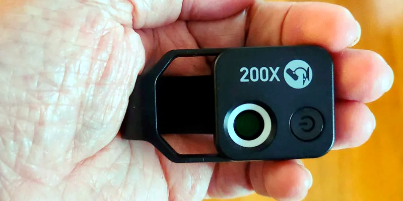
"The researchers designed a fully functional, high-resolution microscope with capabilities close to a modern research microscope. Apart from the optics, all parts were from the toy brick system. The team realized that the lenses in modern smartphone cameras, which cost around €4 each, are of such high quality that they can make it possible to resolve even individual cells."

#biology #microscopy

https://www.uni-goettingen.de/en/3240.html?id=6301